
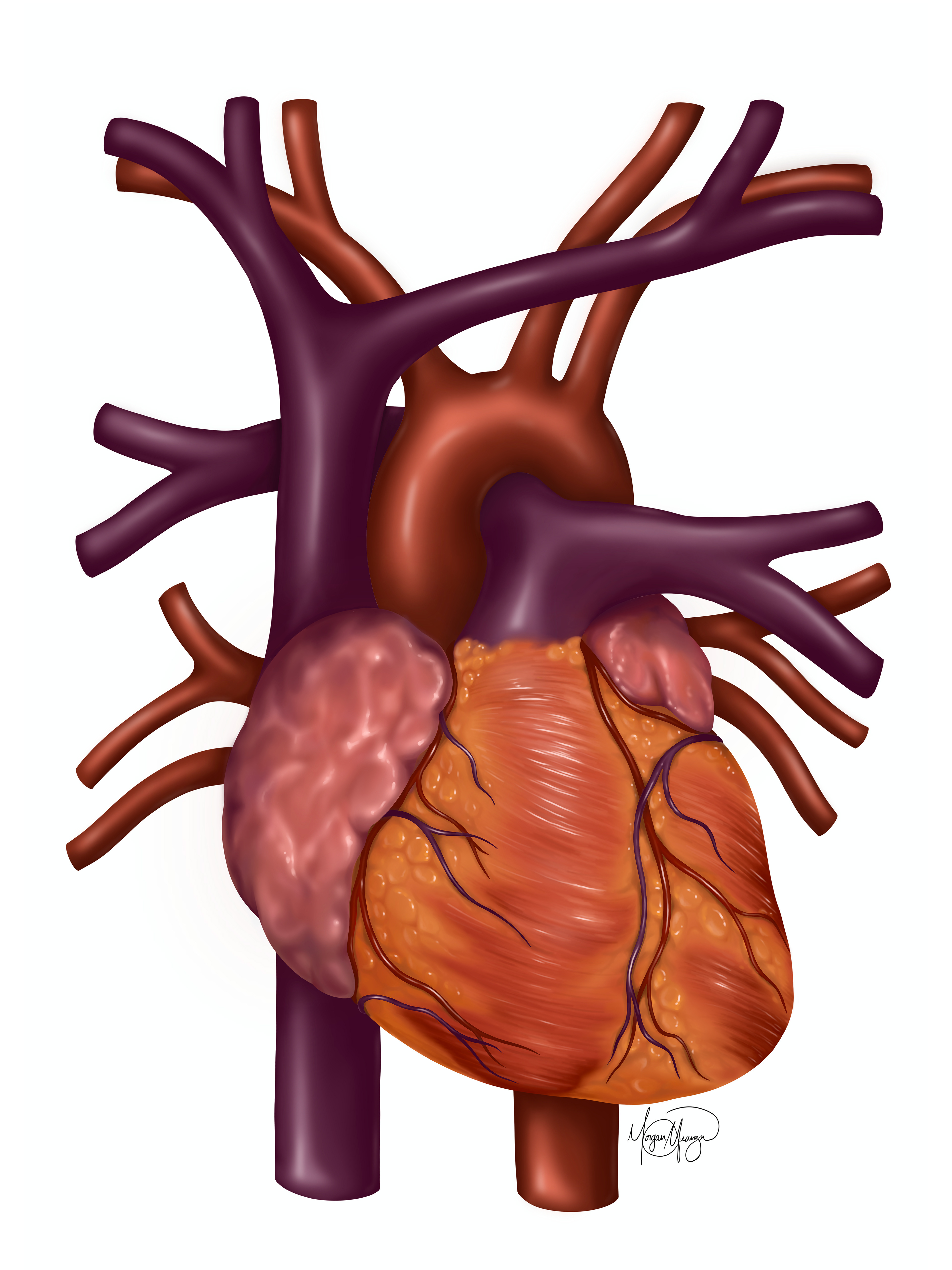
"Healthy Heart" depicts the anterior view of the heart and is left unlabeled to serve a variety of purposes in medical student education. This illustration, alongside many others, are displayed at Ninewells Hospital Cardiac Cath Lab.
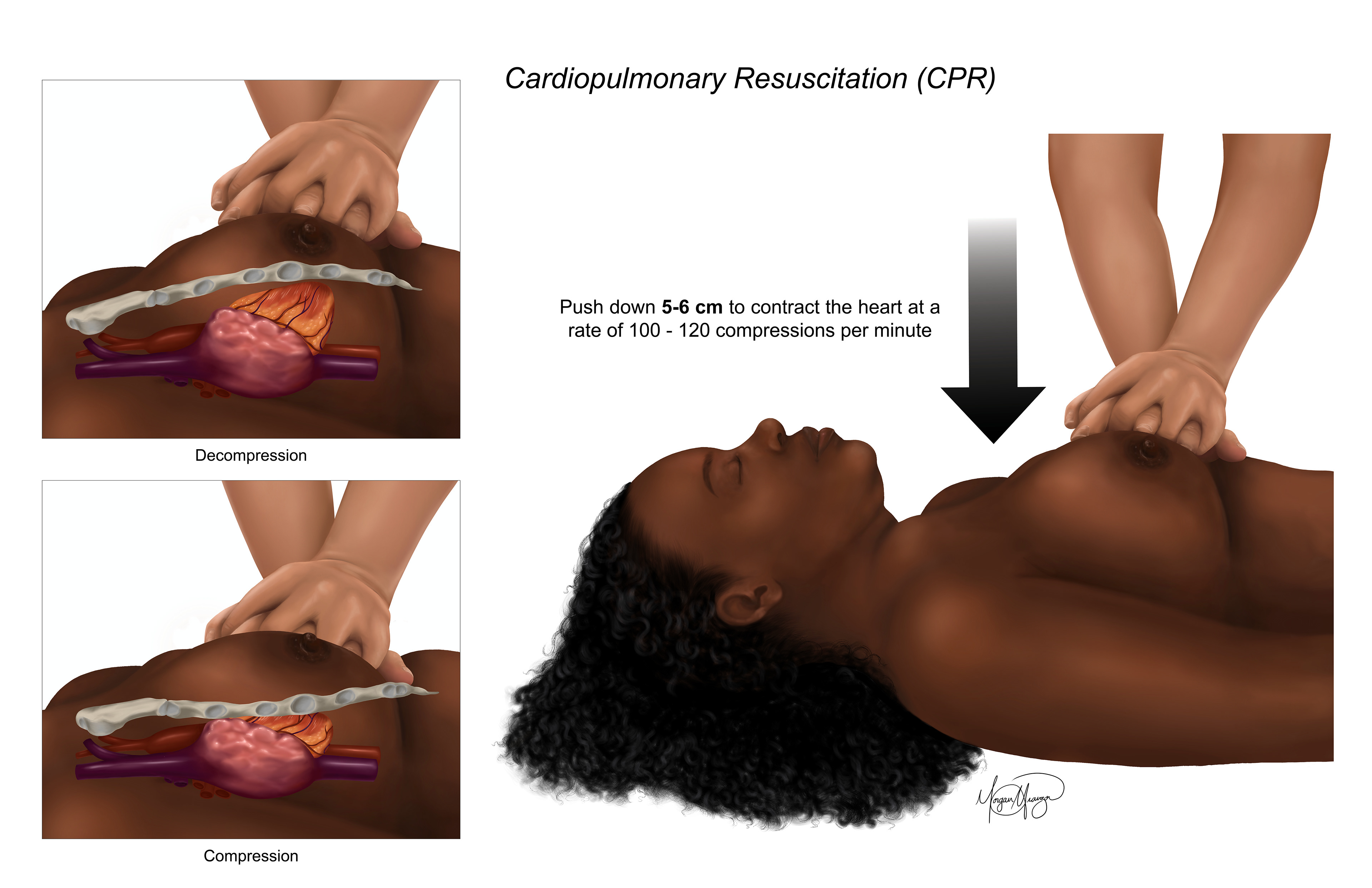
"Cardiopulmonary Resuscitation (CPR)" was designed to be aimed at the general public and encourage people to learn CPR and have more comfort with interacting with women’s bodies during life-saving care. As the research shows, many people are uncomfortable with performing CPR on women due to modesty and fear of touching women’s bodies. By displaying a woman receiving CPR, hopefully viewers will have more comfort in performing life-saving care.
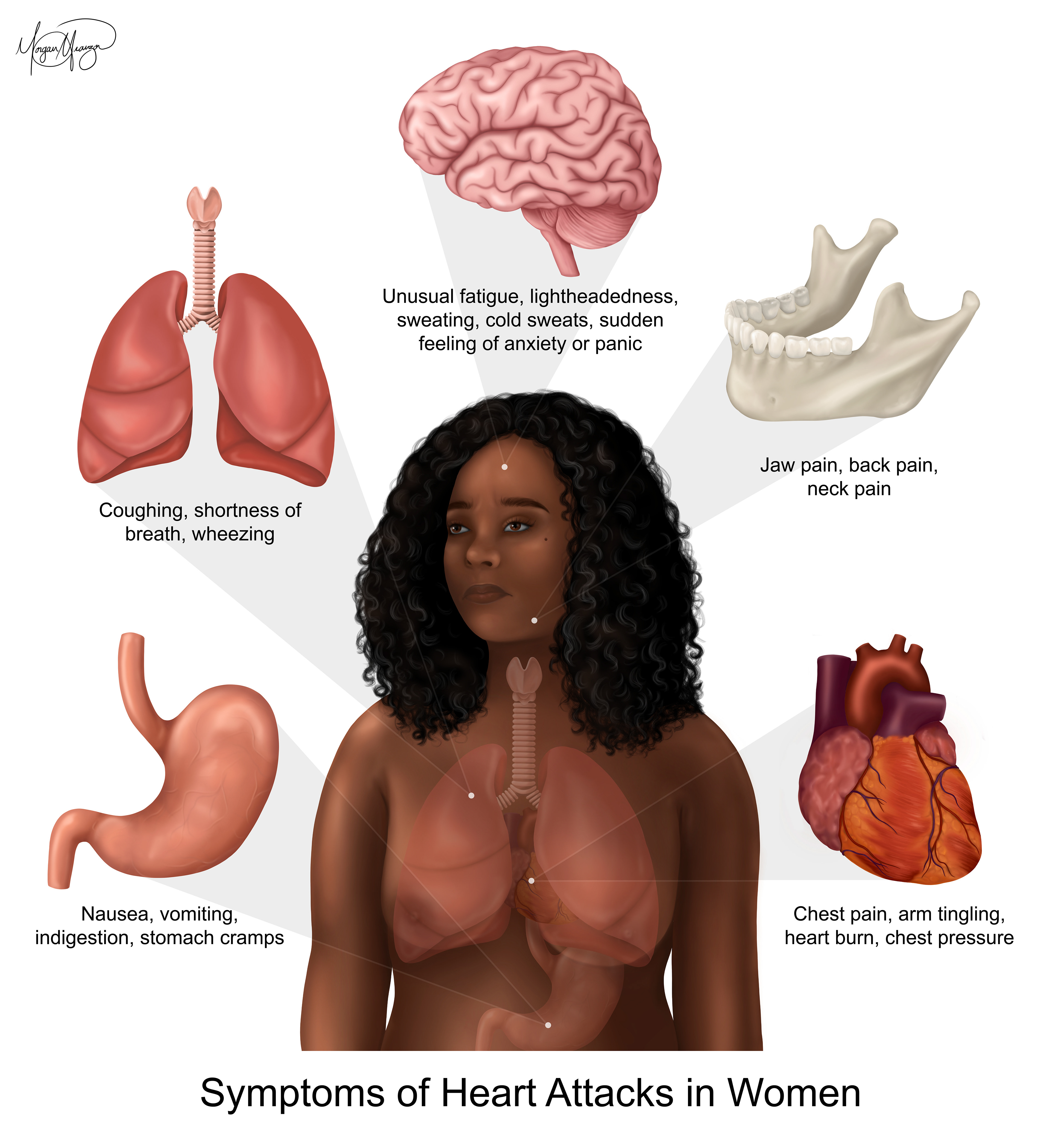
"Symptoms of Heart Attacks in Women" displays symptoms that mainly affect women and have been labeled as atypical, despite affecting half of the population. The intention of this illustration is to educate people on the signs of a heart attack and encourage individuals to seek medical care if they are experiencing these symptoms.
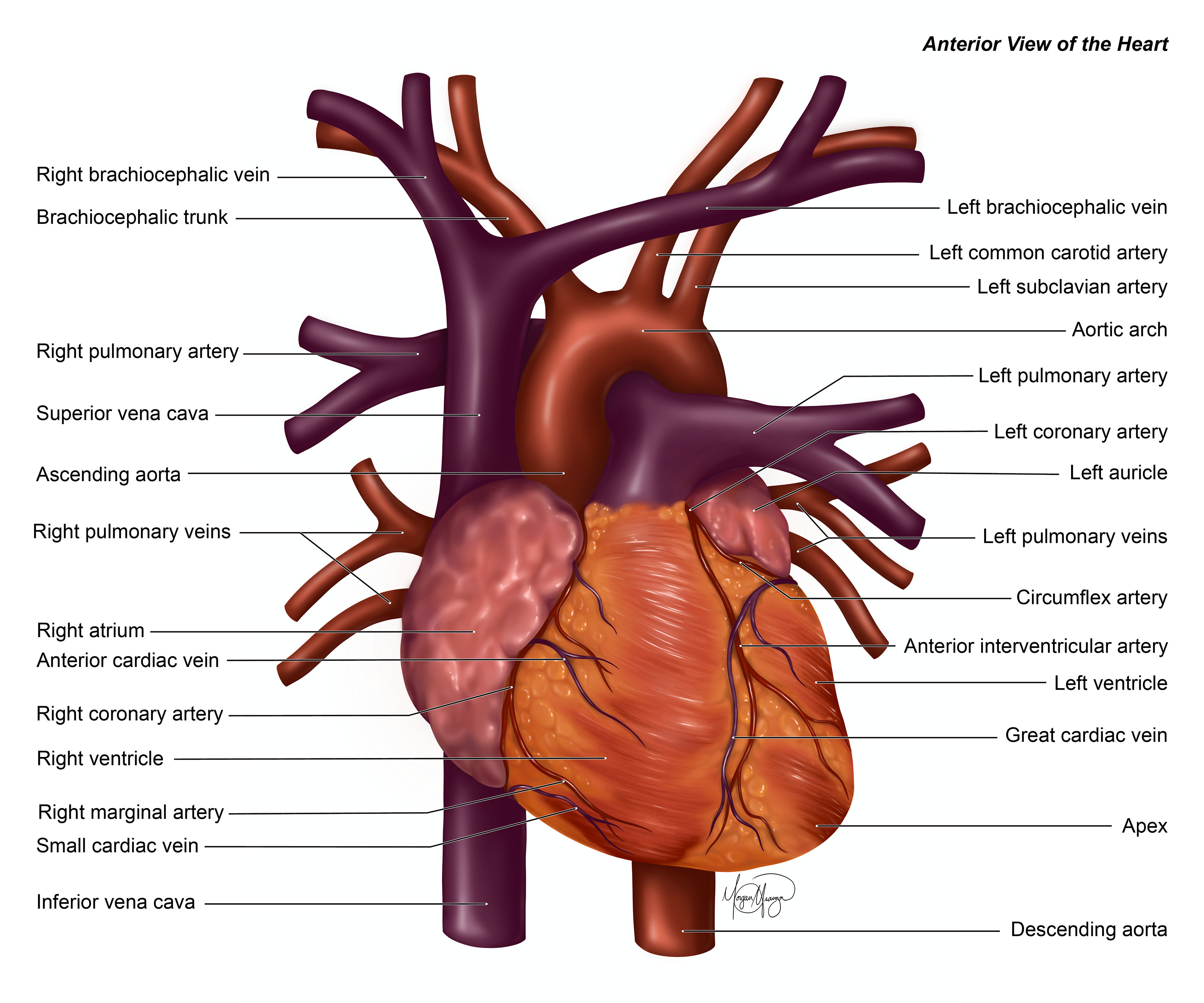
"Anterior View of the Heart" was created for individuals to learn about the heart anatomy. This diagram labels the anterior anatomy of the heart for medical students learning about the heart or patients to follow in conversations with their doctors.
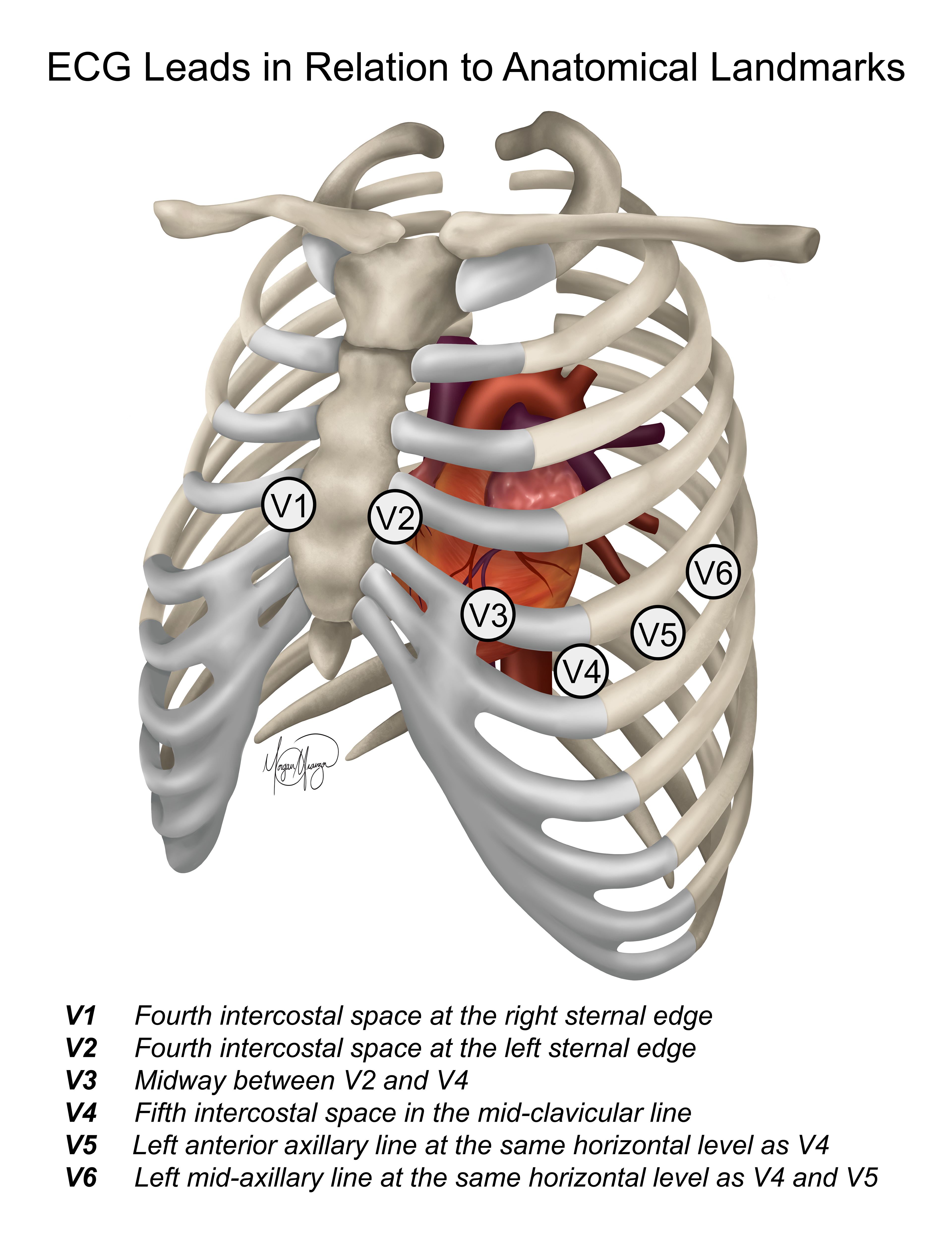
"ECG Leads in Relation to Anatomical Landmarks" showcases the landmarks medical professionals use when placing the chest leads. While all body types vary, the underlying anatomy is the same, thus creating an “anatomical” guide for where the leads go. This illustration visualizes where those leads are aiming for when placed on the skin.
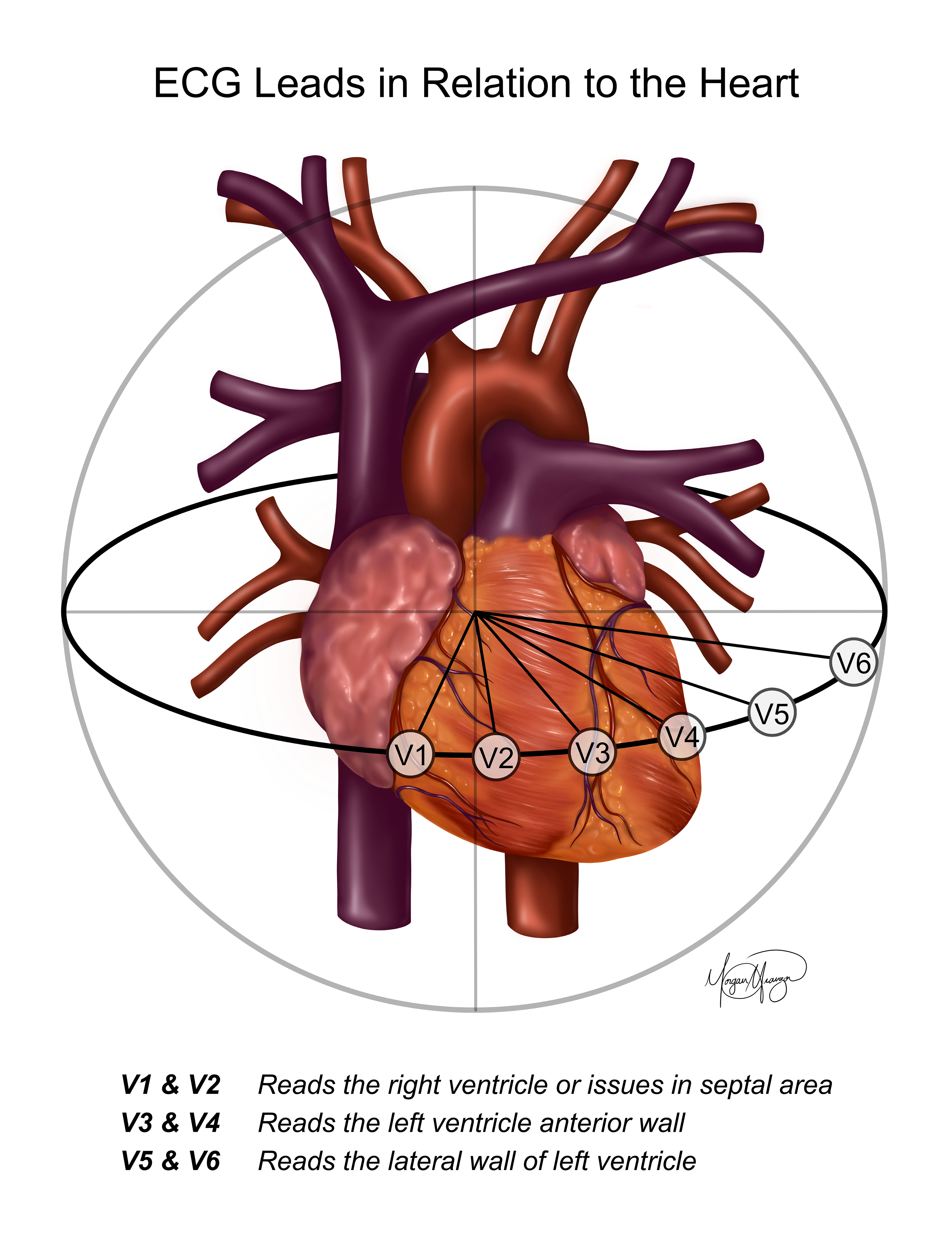
"ECG Leads in Relation to the Heart" visualizes how to read the ECG results. Each precordial lead corresponds to different areas of the heart, allowing medical professionals to locate which area of the heart is not working properly.
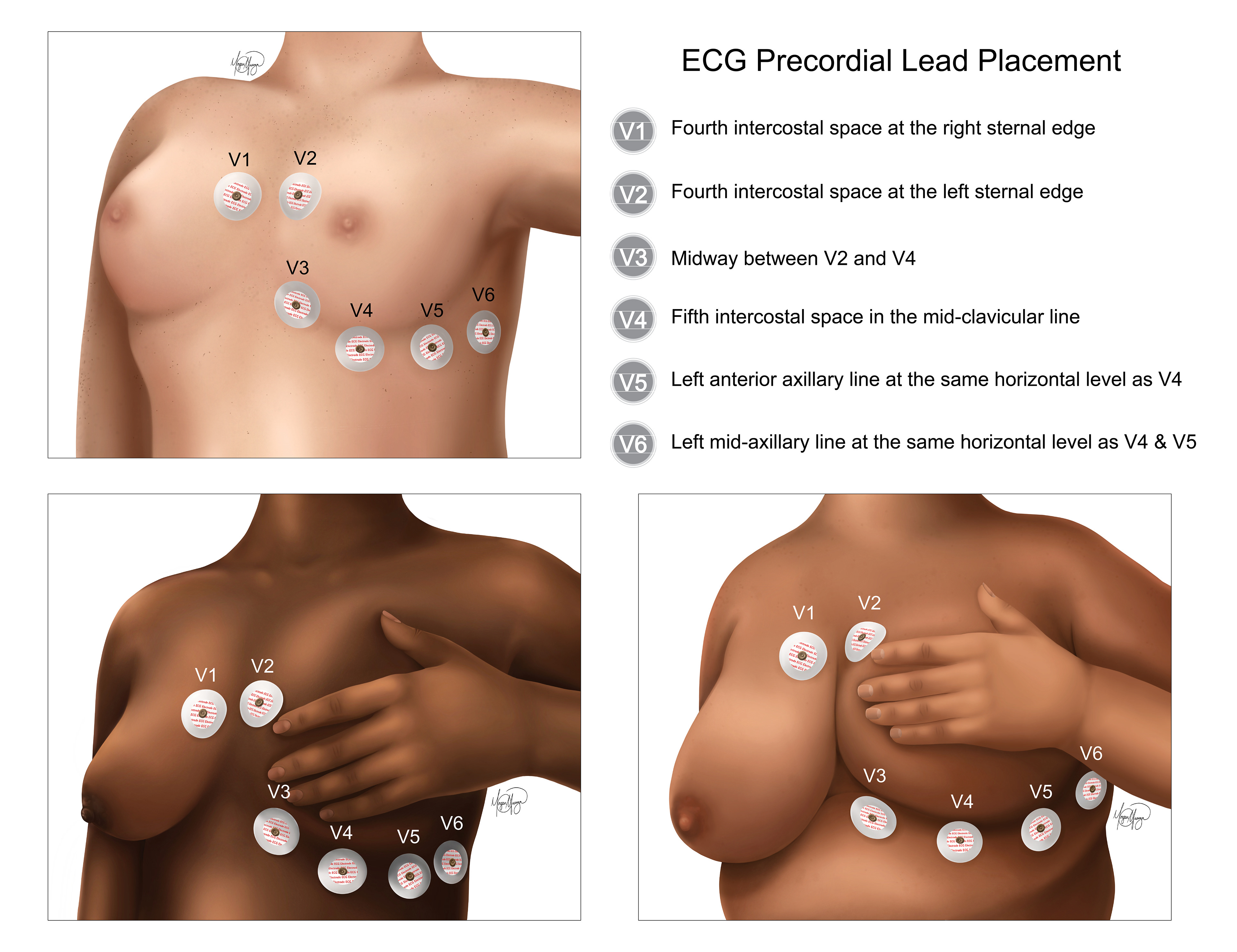
"ECG Precordial Lead Placement" consists of three illustrations of different body types and skin tones. This illustration is incredibly important because incorrect lead placement can result in misinterpretation of the ECG and electrical interference. In order to avoid this, medical professionals are asked to lift the breast, or have patients lift their breast, and place the leads correctly.
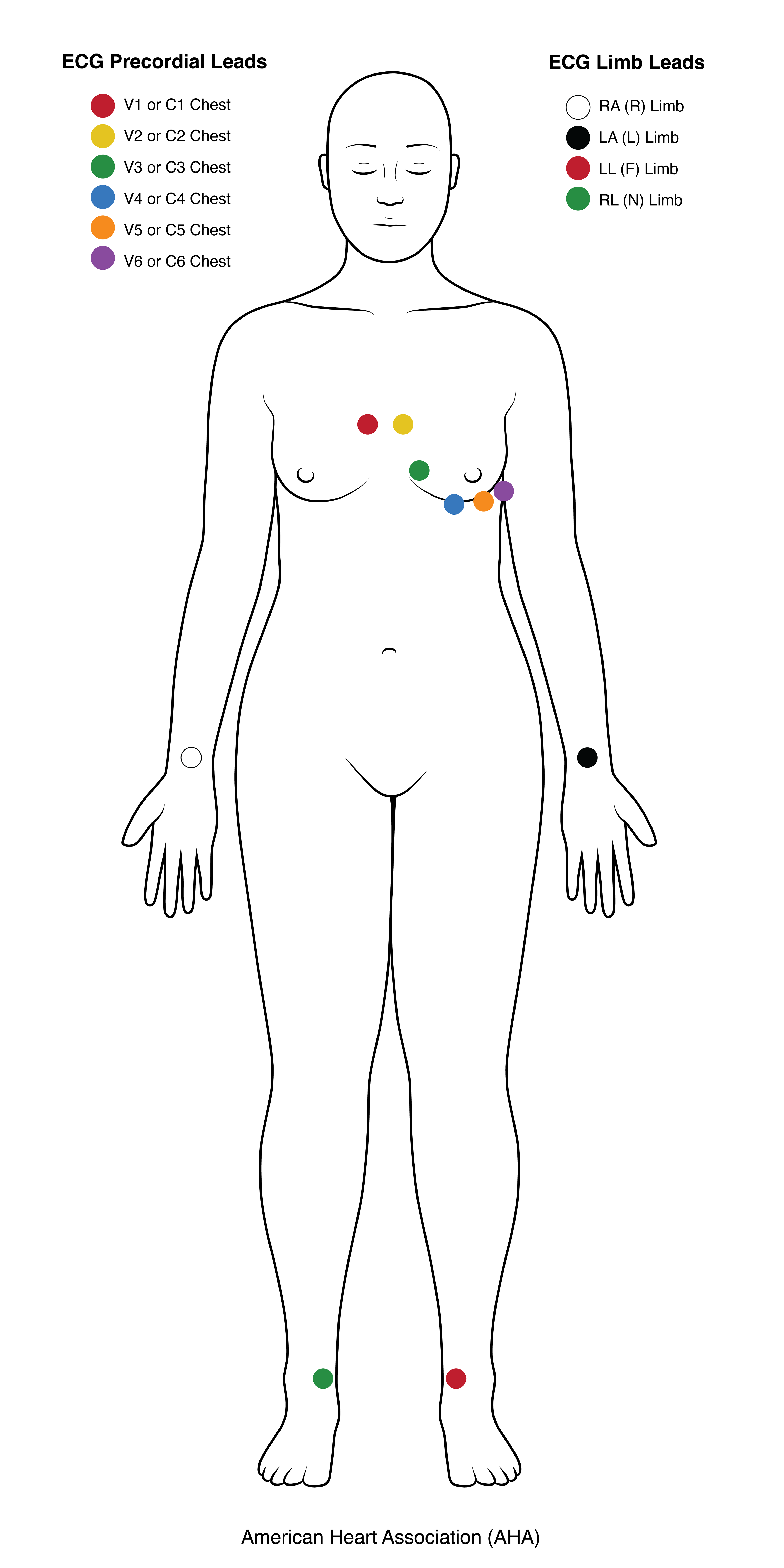
This additional illustration goes alongside "ECG Precordial Lead Placement" to showcase the limb leads, as well.
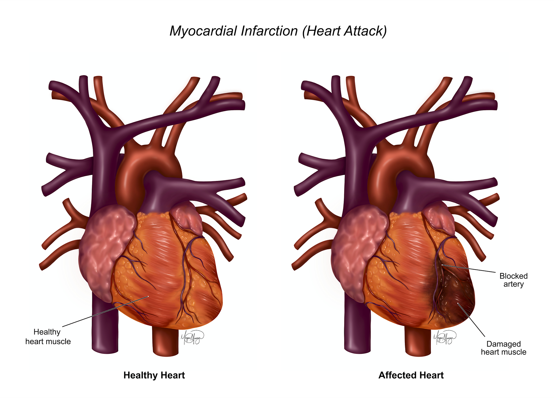
"Myocardial Infarction (Heart Attack)" compares a healthy heart to one affected by a heart attack. While most individuals have heard of a heart attack, they might not understand what exactly is happening inside the body. This piece visualizes the anatomical differences when an artery is blocked and affects the muscle of the heart.
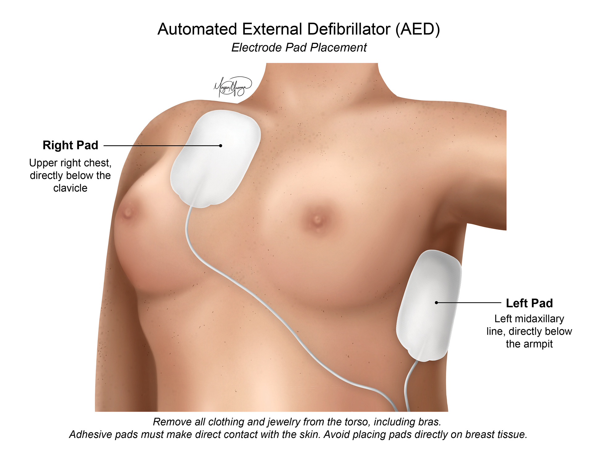
"Automated External Defibrillator (AED)" depicts the proper placement of a defibrillator, targeted at the general public. AEDs are often in most public settings and come with basic instructions, meaning that anyone can use one without any prior experience. This illustration is aimed at providing a visual to follow, while also providing reassurance that removing all clothing and cutting off a woman’s bra is completely necessary.