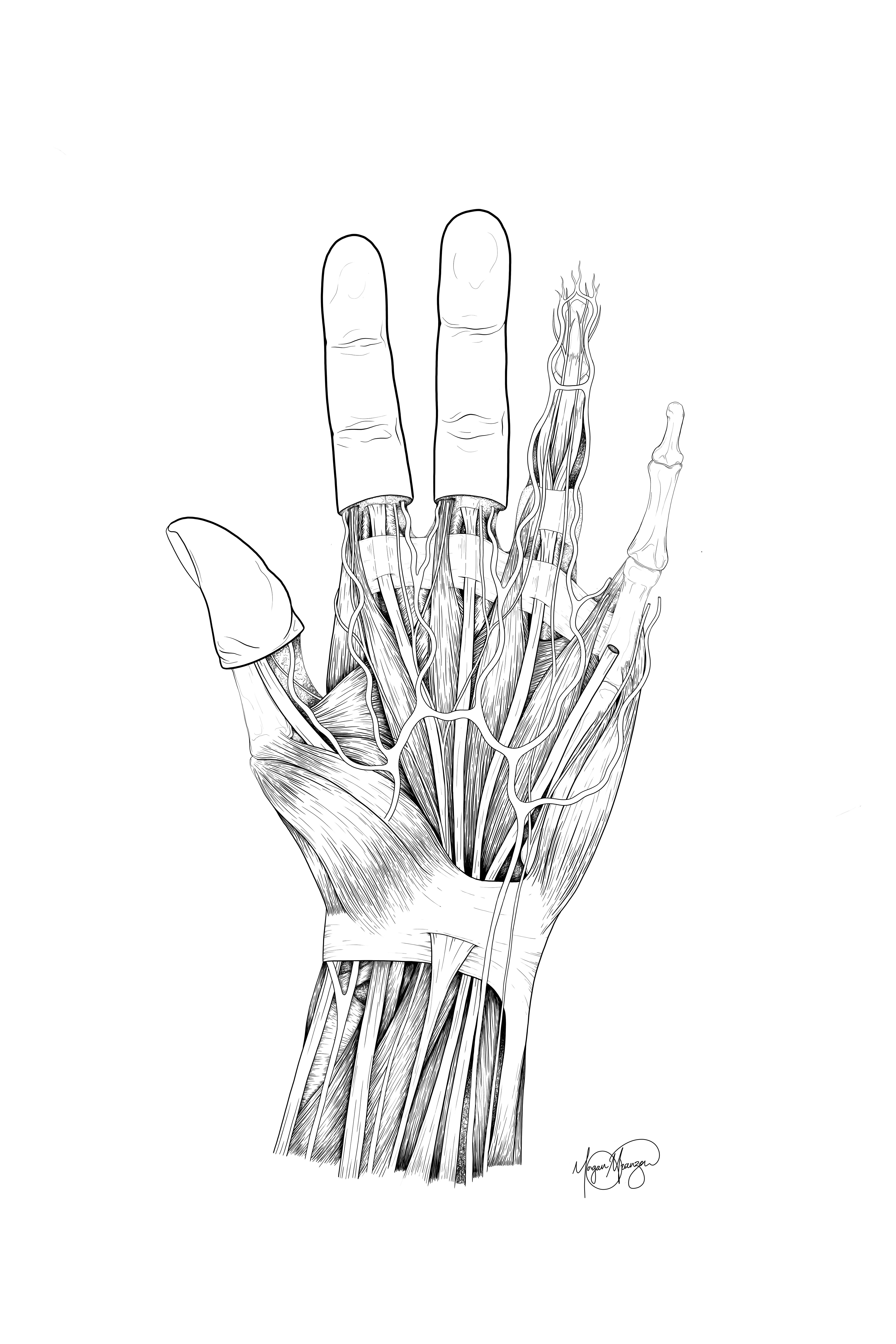
Anterior dissection drawing of the hand, comparing a typical hand to one with hypermobility. The hypermobility is shown through hyperextension of the metacarpophalangeal joint with some flexion at the carpometacarpal joint. In patients with hEDS or hypermobility, joints have a higher range of motion as a result of weak muscle and tendon support at the joint, causing discomfort, fatigue, and instability that can cause longterm issues.
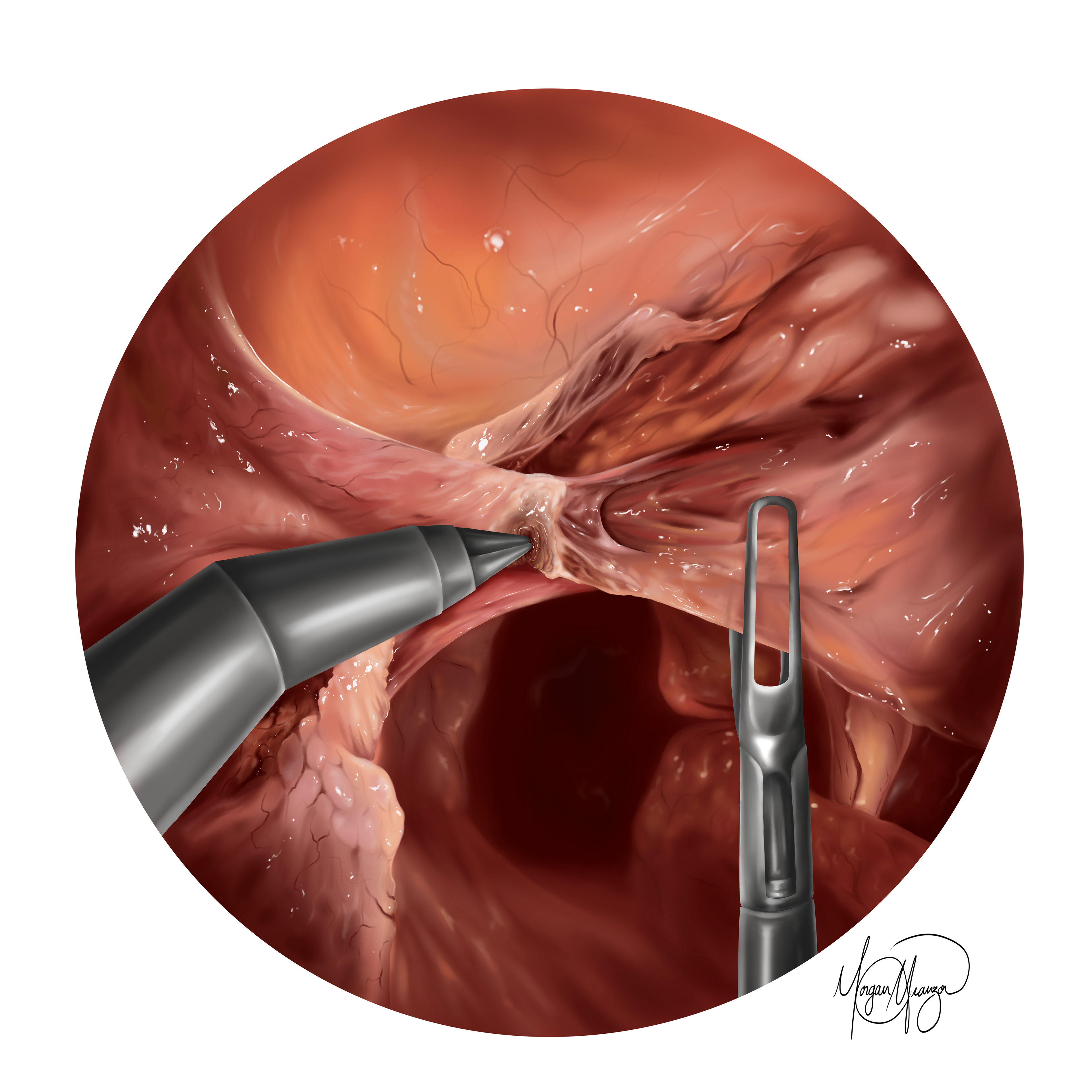
Surgical illustration of a total laparoscopic hysterectomy, part of a series of 5 illustrations. This visual depicts the ligation of the uterine artery (representing multiple steps in the procedure of cauterizing surrounding blood vessels).
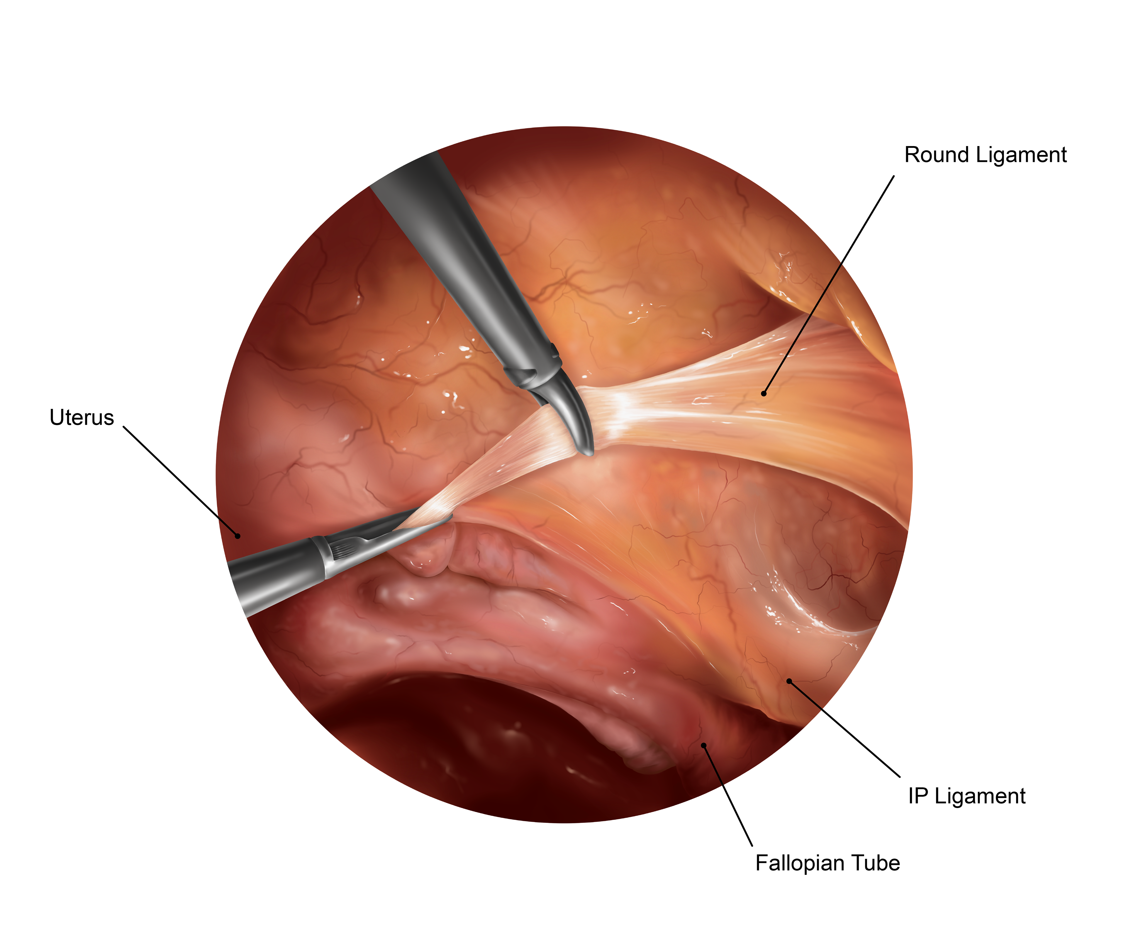
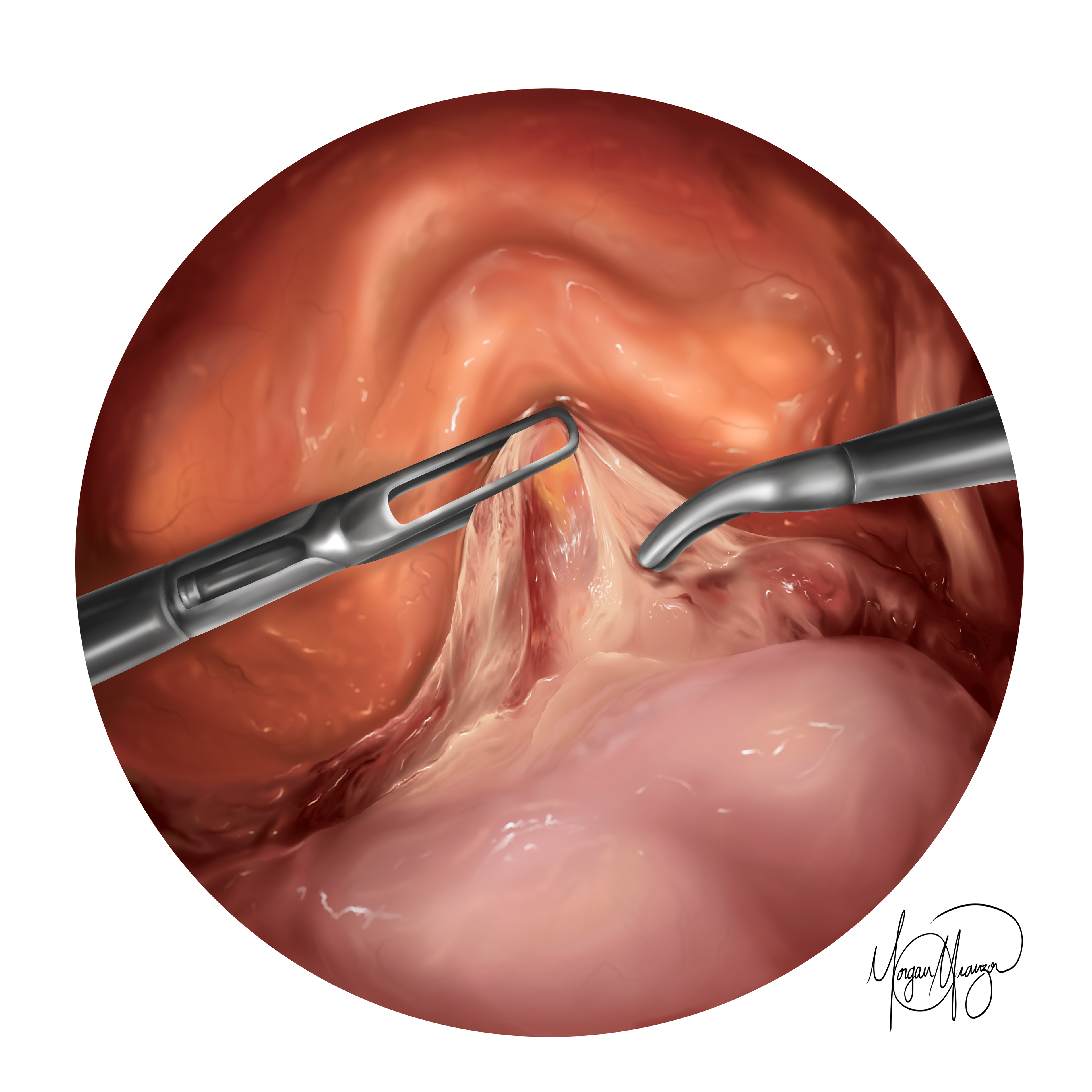
Surgical illustration of a total laparoscopic hysterectomy, part of a series of 5 illustrations. This labeled visual displays the transection of the round ligament (representing various steps to divide all the supporting ligaments).
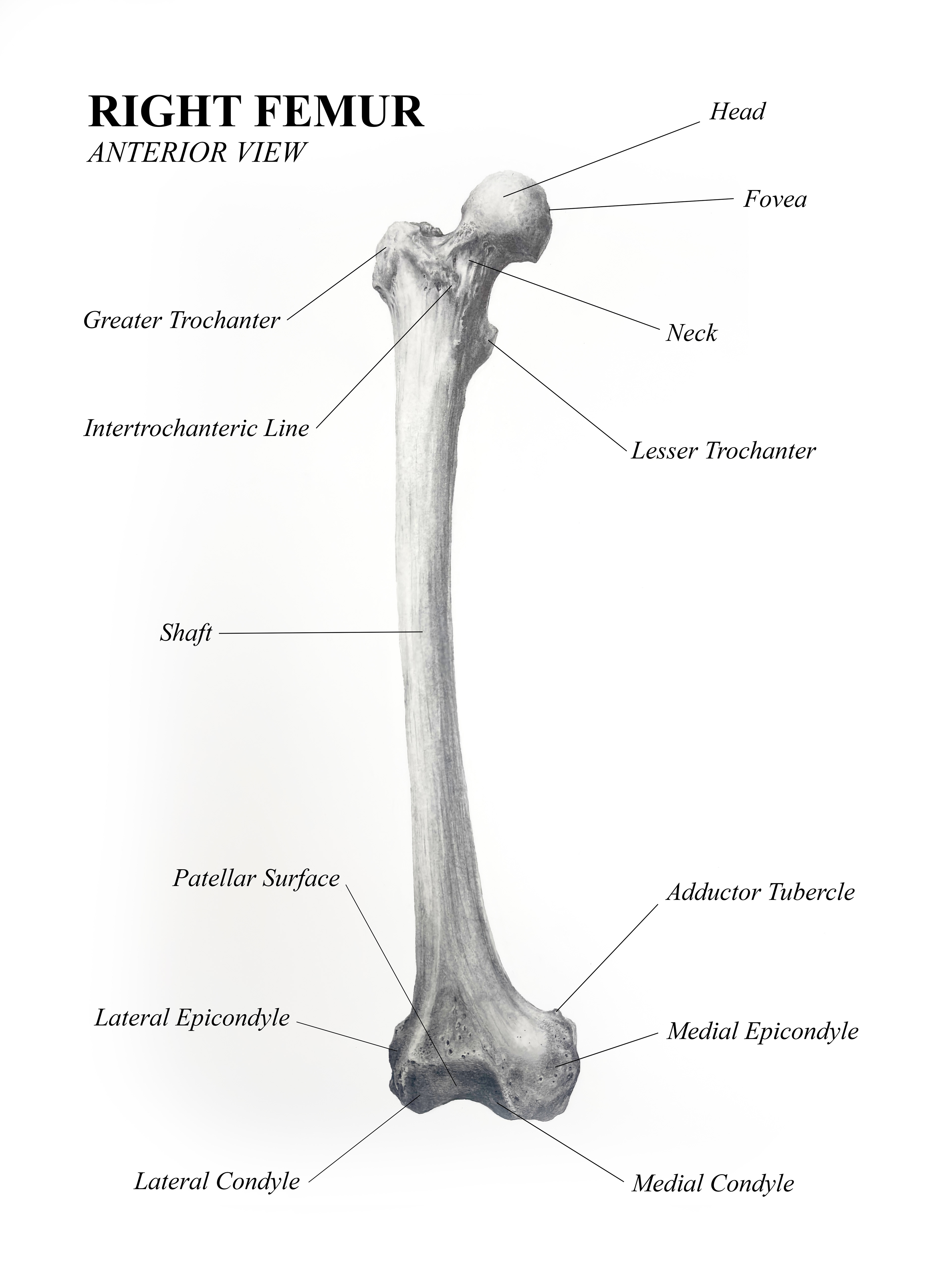
Anterior Femur Diagram (Carbon Dust and InDesign, 2025)
Total Laparoscopic Hysterectomy. b (Procreate, 2026)
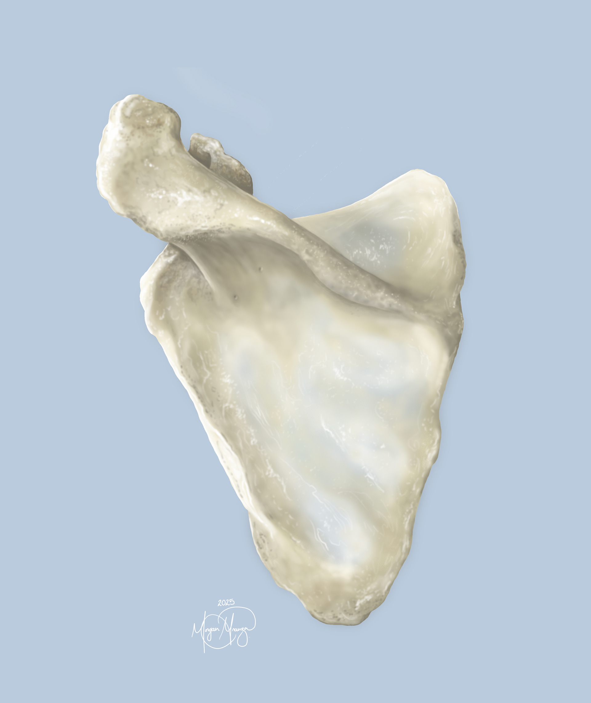
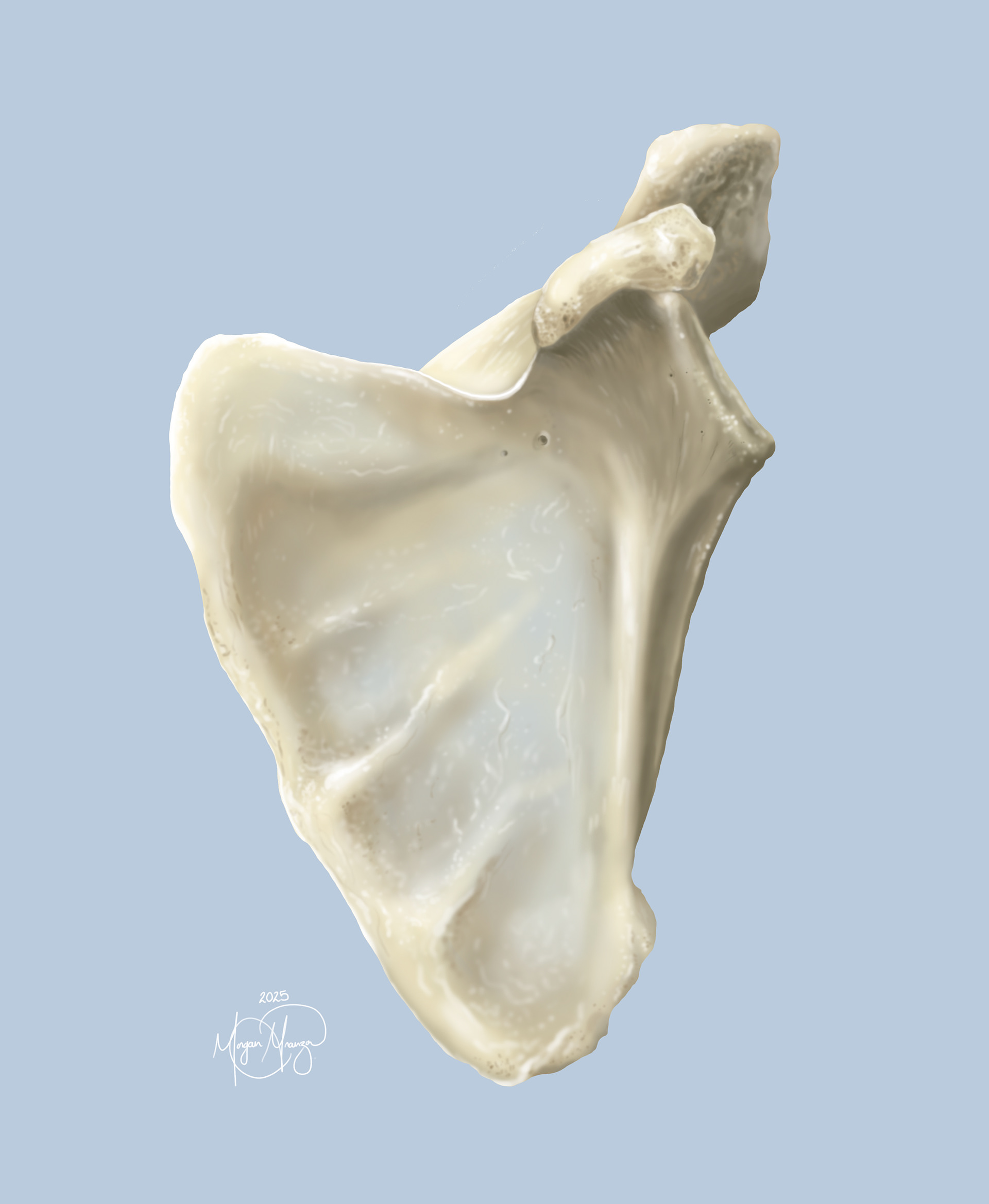
Anterior Scapula (Photoshop, 2025)
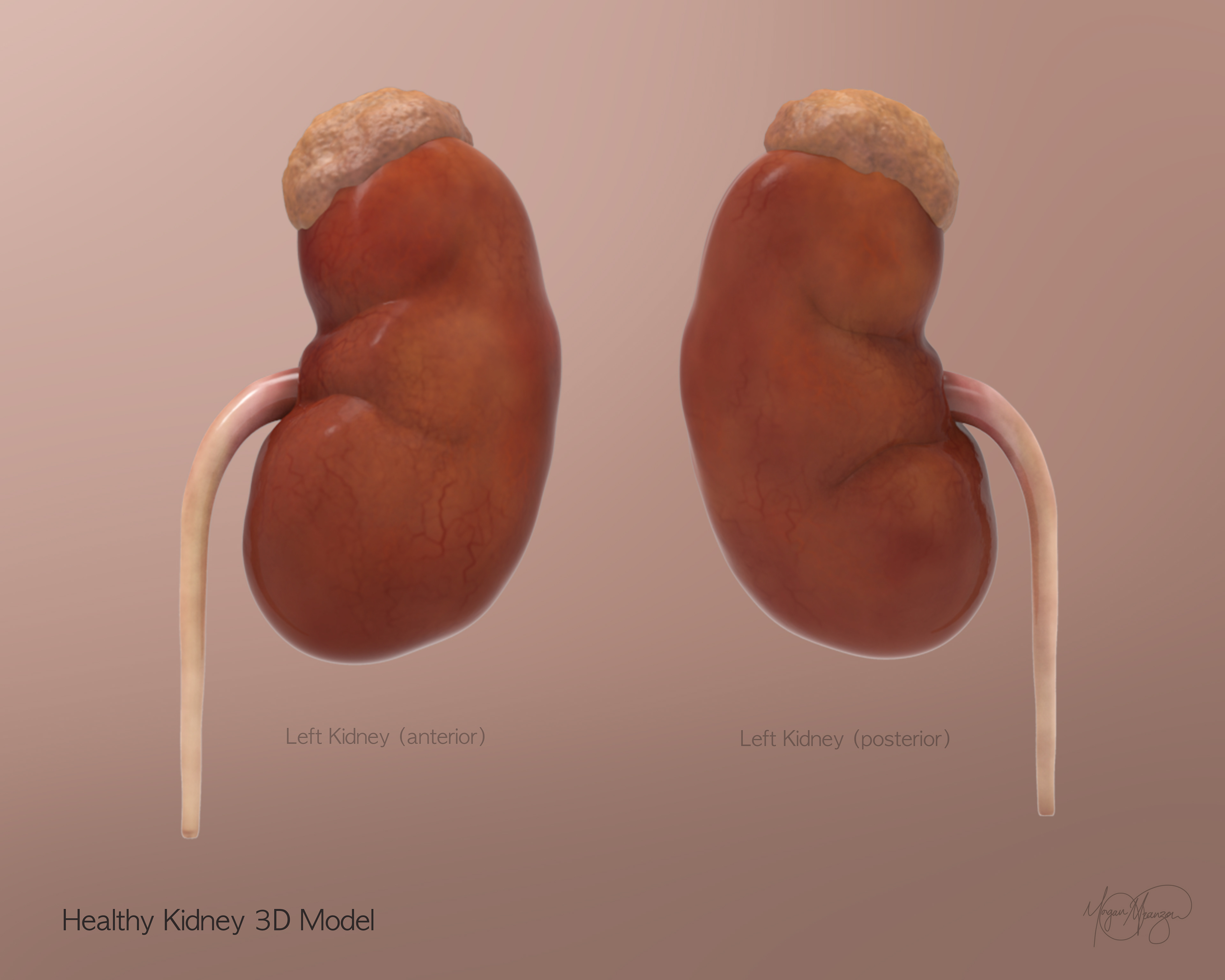
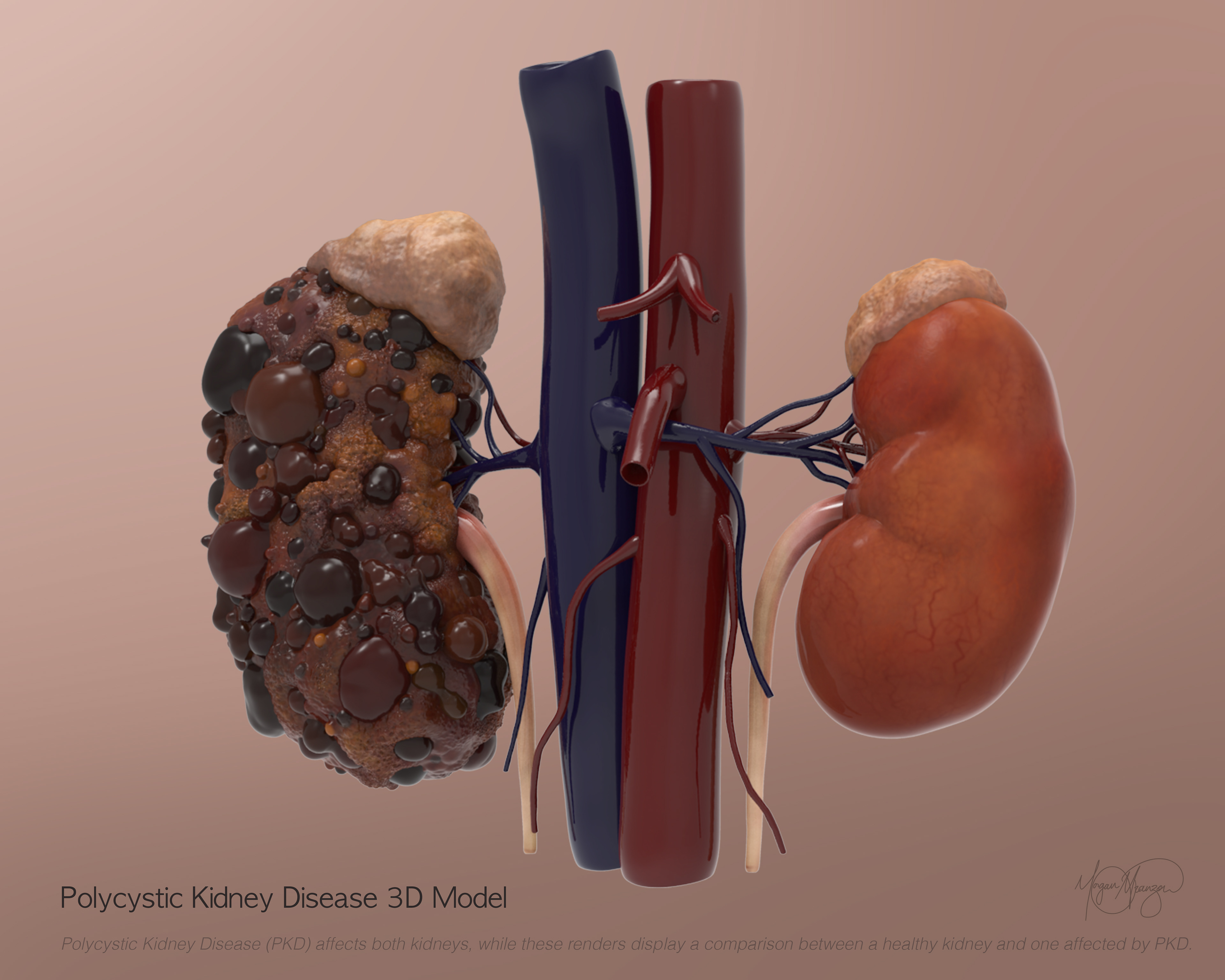
Healthy Kidney 3D Model (ZBrush, 2026)
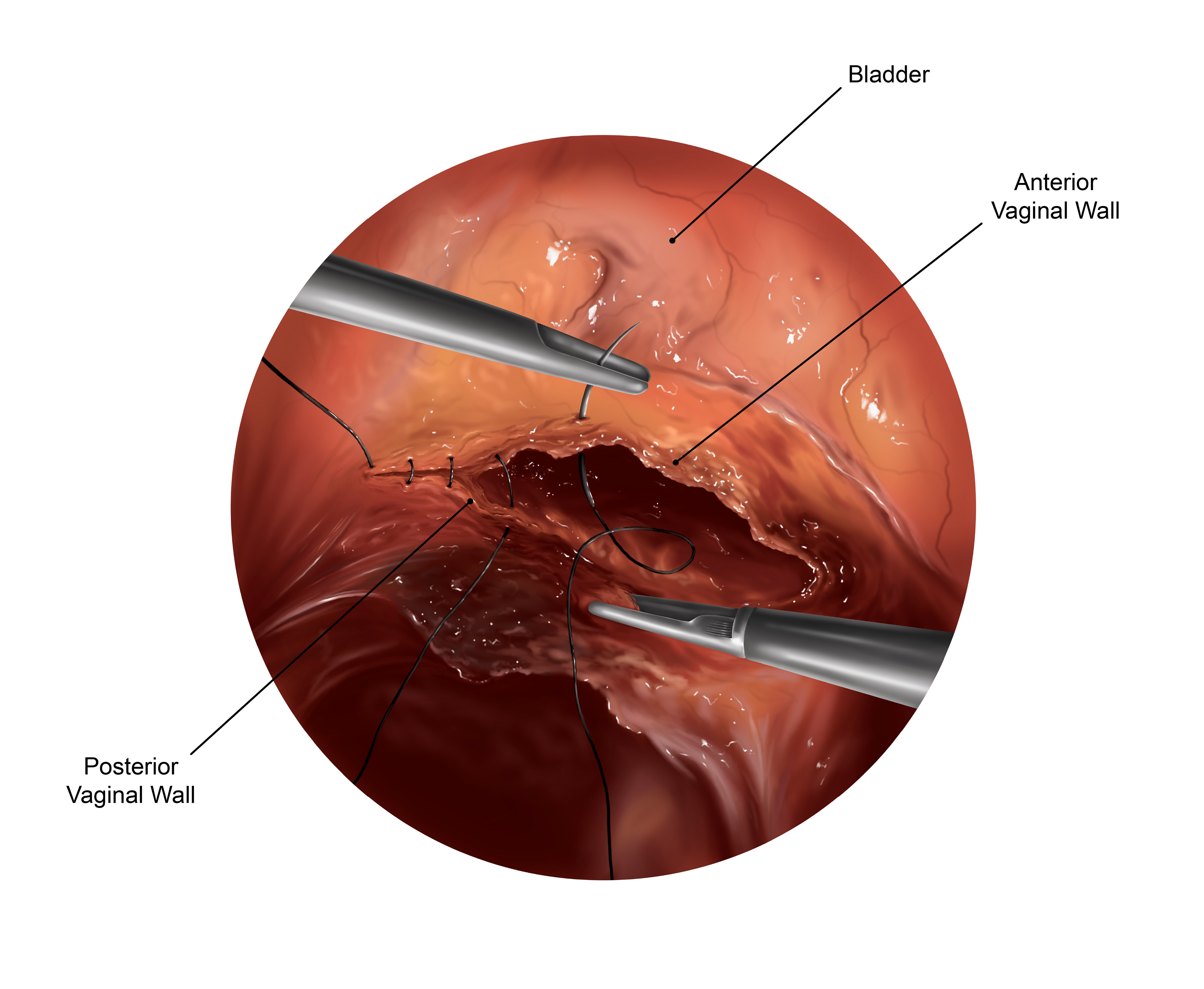
Total Laparoscopic Hysterectomy. e (Procreate, 2026)
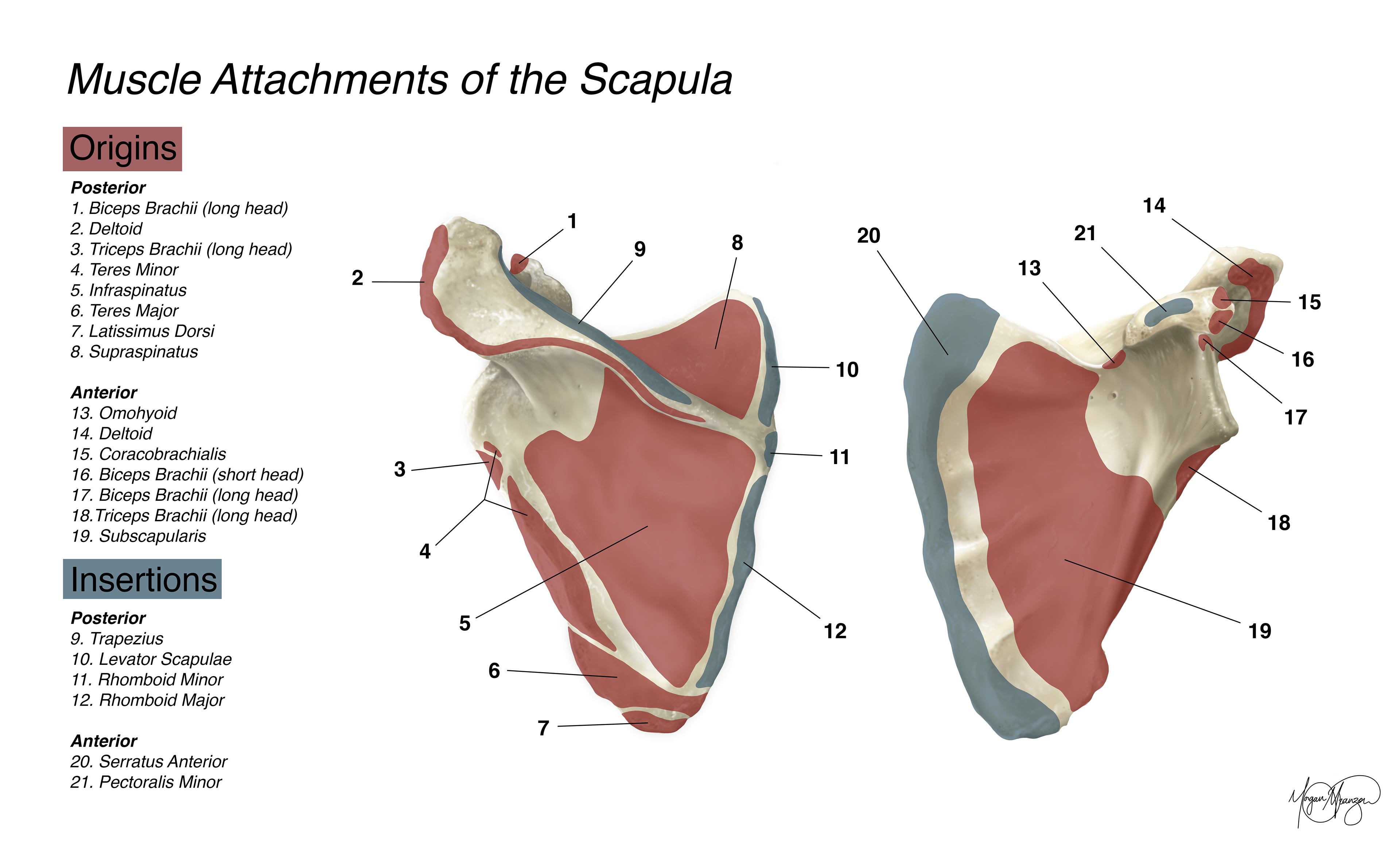
Muscle Attachments of the Scapula (Photoshop, 2025)
Posterior Scapula (Photoshop, 2025)
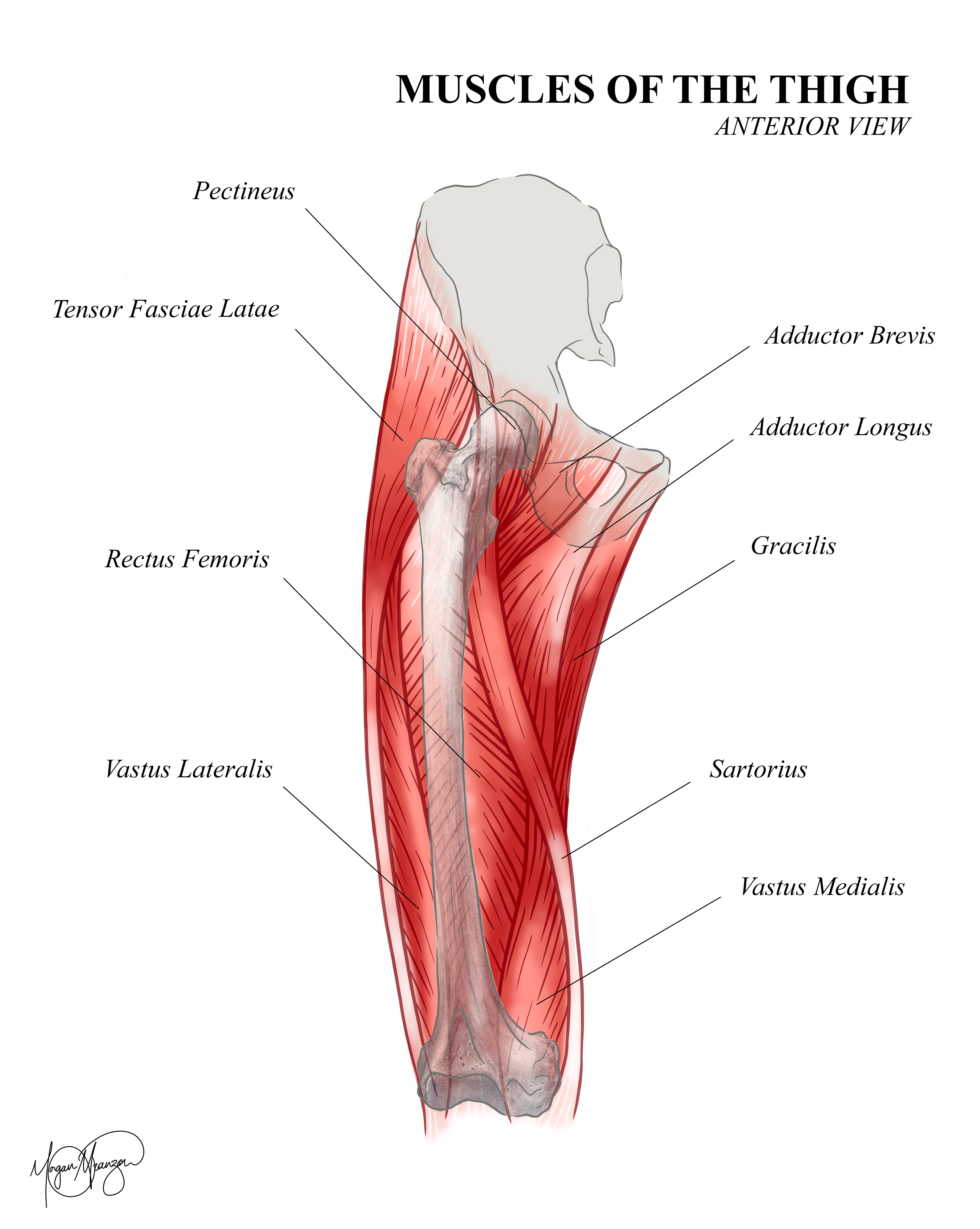
Muscles of the Thigh (Photoshop, 2025)
Hypermobile Dissection of the Hand (Photoshop, 2025)
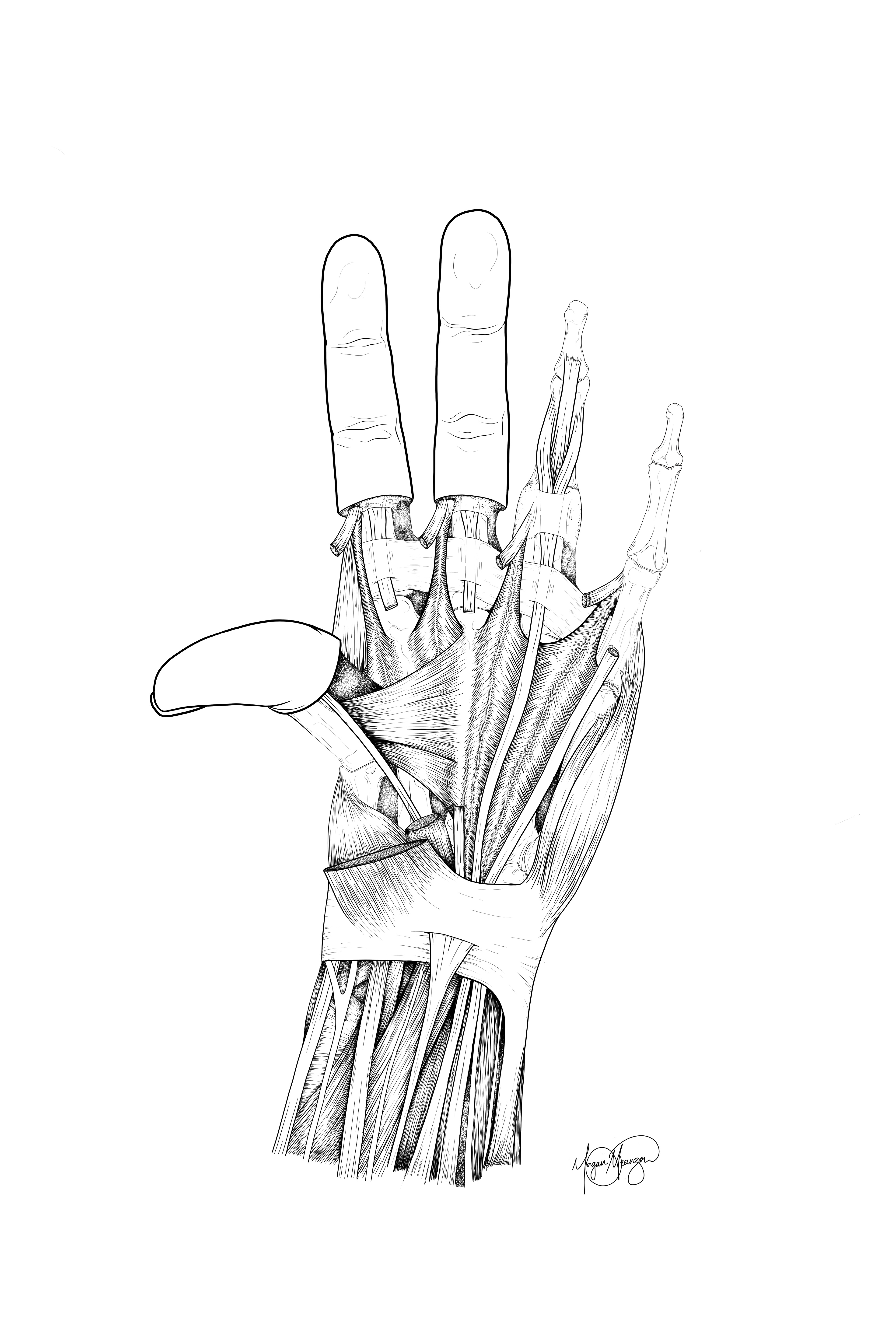
Anterior Dissection of the Hand (Photoshop, 2025)
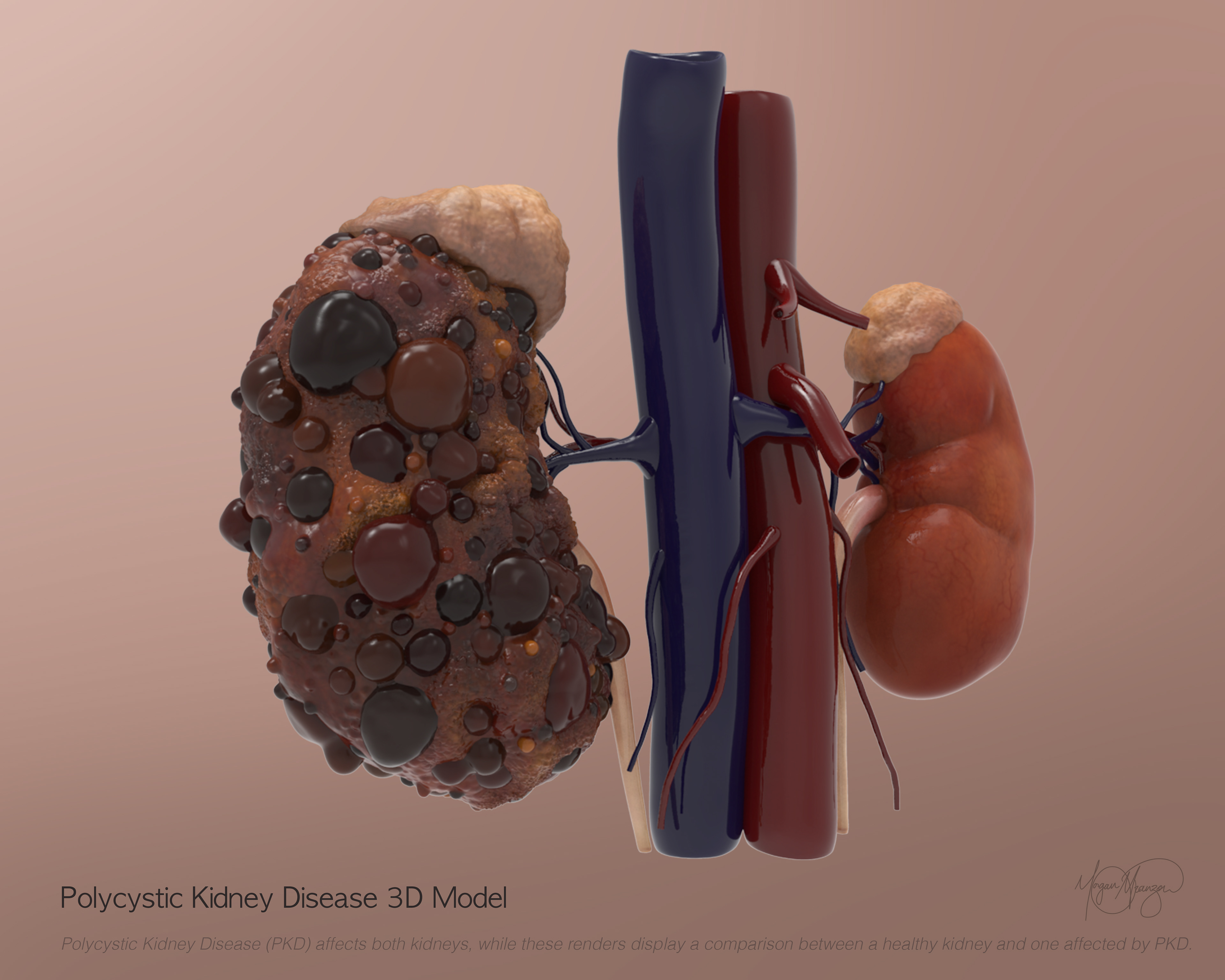
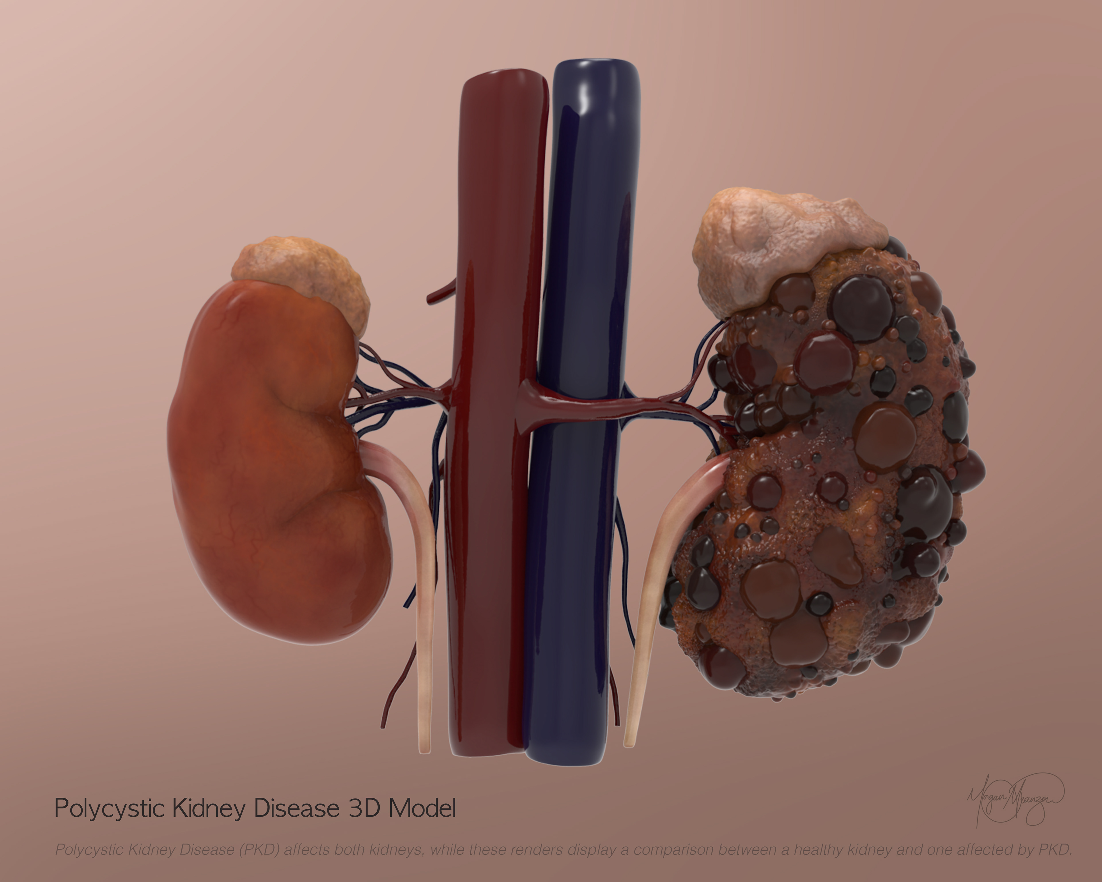
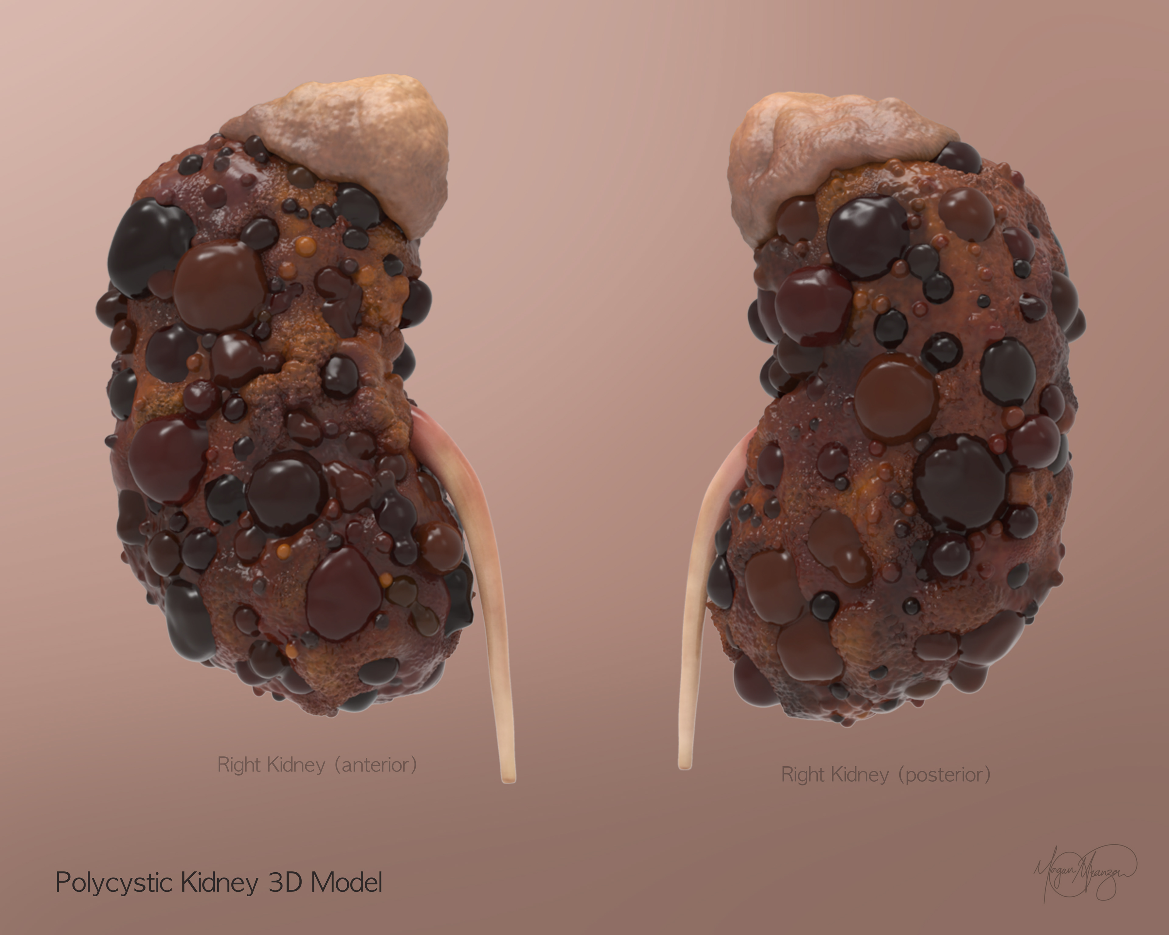
Polycystic Kidney 3D Modle (ZBrush, 2026)
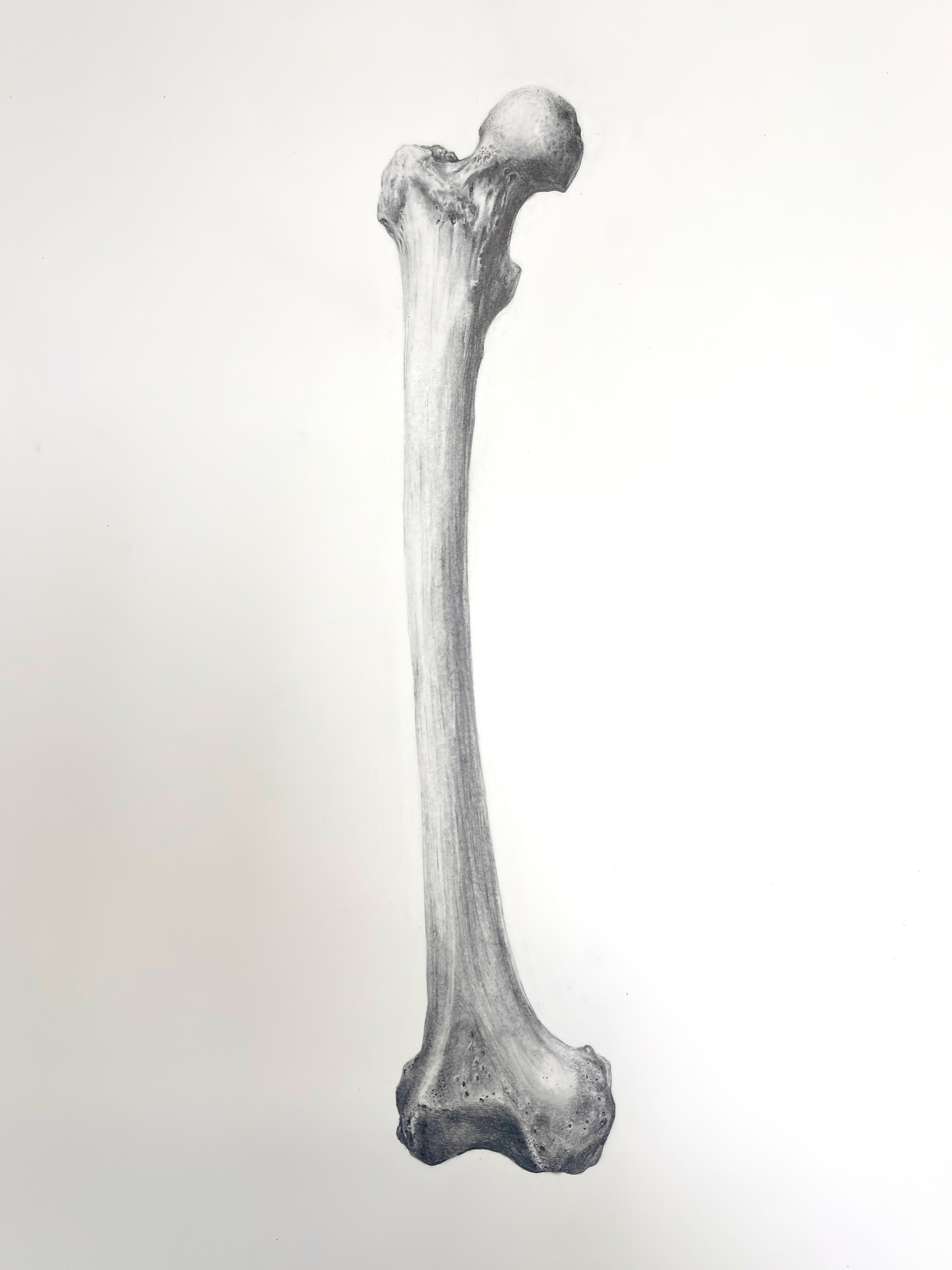
Right Femur (Carbon Dust and Graphite Pencil, 2025)
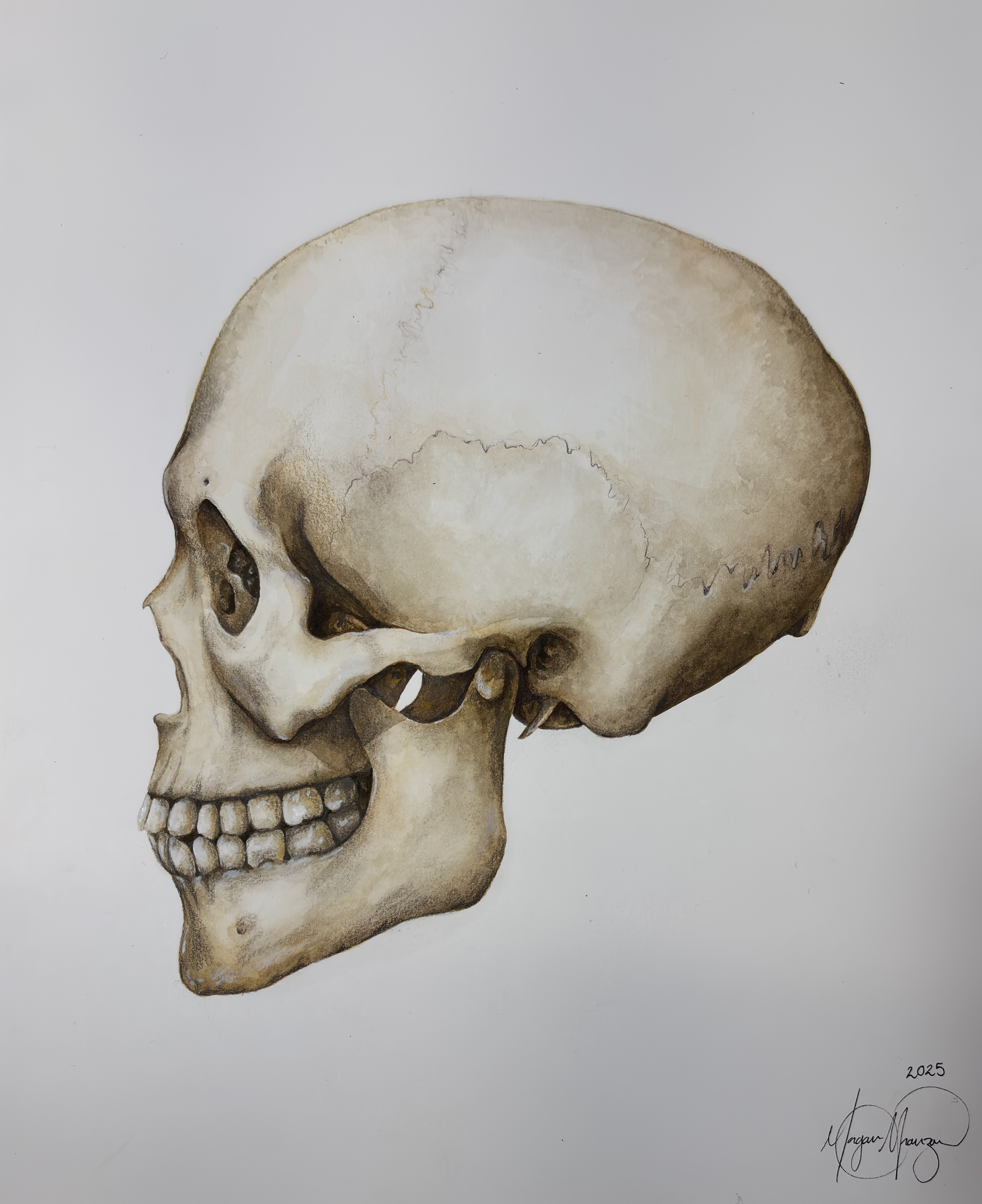
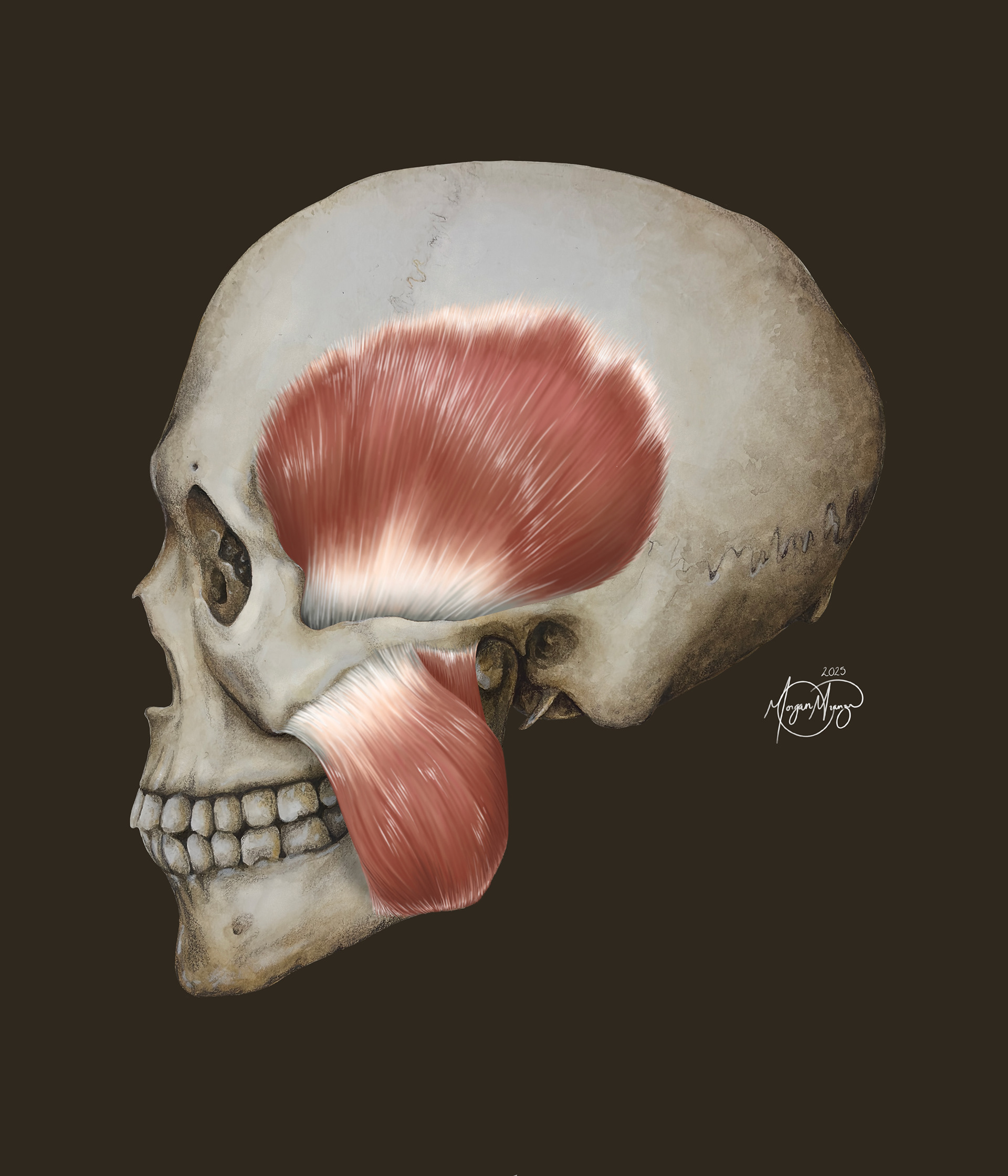
Watercolor Skull (Watercolor, 2025)
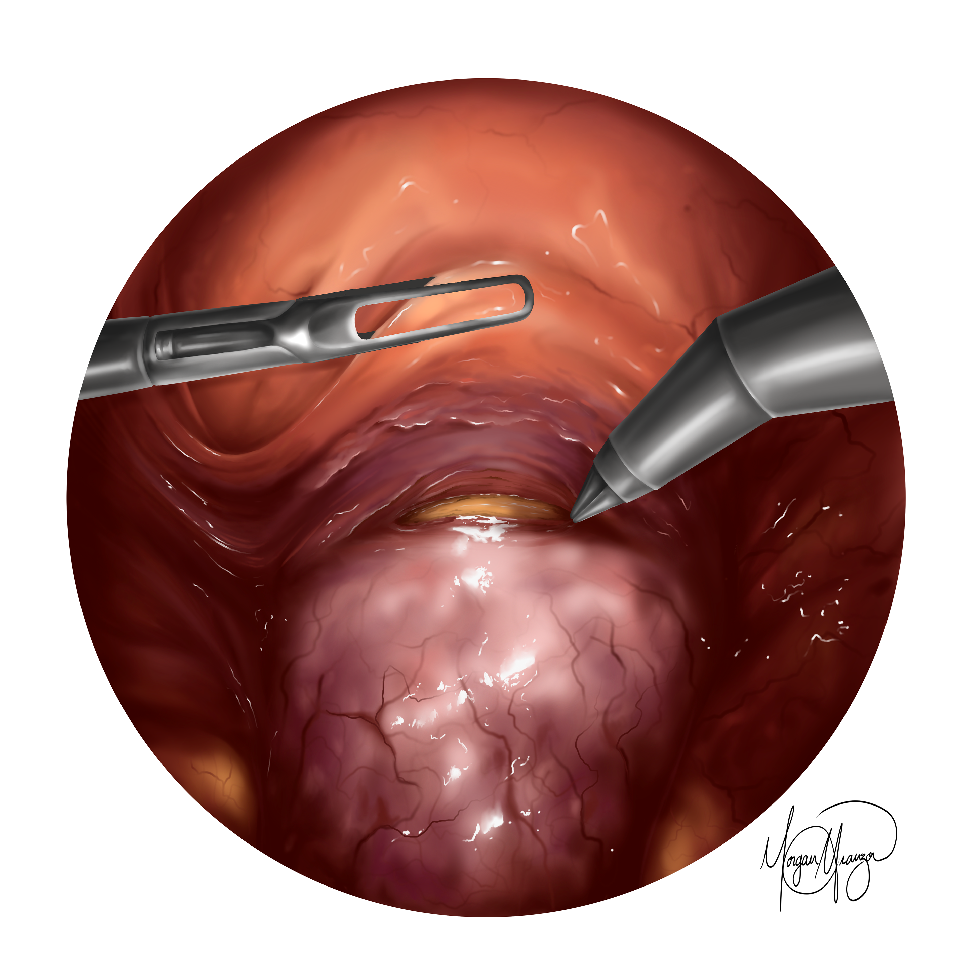
Total Laparoscopic Hysterectomy. d (Procreate, 2026)